Stay up to date with what the London & Kent Bone & Joint team are up to…
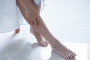
Lower Limb
![]()
The knee joint is made up of bones, ligaments and tendons. The knee itself is a modified hinge joint and joins the thigh bone (femur) and the shin bone (tibia). The kneecap (patella) and the fibula (smaller shin bone) are the other bones which comprise the knee joint. Ligaments such as the medial and lateral collateral ligaments as well as the anterior and posterior cruciate ligaments help to stabilise the knee joint in each direction. The muscles and tendons (which join the muscles to the bones) also help with stability as well as movement of the knee joint.
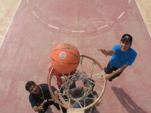
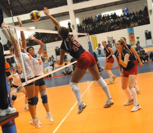
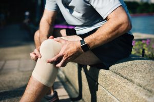
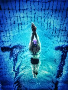
Knee tendonitis, or patellar tendonitis, is an overuse injury of the tendon which connects the kneecap to the shin bone. This tendon, along with the muscles at the front of the leg and knee, allow knee extension for movements like running, jumping and kicking. This injury is often seen in athletes who are required to do lots of jumping as part of their sport.
Knee tendonitis symptoms usually start with pain, specifically between the kneecap and there the tendon joins the tibia. This pain can occur when doing physical activity, get worse when playing sport or eventually become so painful that it inhibits daily activities like climbing the stairs. The pain can be exacerbated when playing sport or performing regular movements because it is an overuse injury meaning it is caused by repetitive stress on the tendon. The pain is a result of small tears in the tendon which the body automatically tries to repair. As the tears increase in number, the natural reaction of the body is pain and inflammation in the area as well as weakening of the tendon. Ultimately, continuing to play sport or perform the activity through the pain will make the condition worse.
There are some factors which can predispose an individual to knee tendonitis which include:
- Physical activity – specifically jumping and running as these increase the stresses on the tendon.
- Muscle imbalance – if some muscles are stronger than other these can pull on the tendon, dragging it out of line.
- Tight leg muscles – the hamstring and quadriceps muscles can put strain on the tendon if they are very tight.
The general rule is that if the pain in the knee following physical activity is not alleviated with rest or ice, it is a good idea to see a doctor. The little tears that have formed and are the root cause of the pain will continue to get worse if left untreated so if you’re worried, it’s always a good idea to seek advice. Without the correct treatment, the pain and swelling can continue to get worse and could lead to a much worse condition like tendinopathy.
This article is intended to inform and give insight but not treat, diagnose or replace the advice of a doctor. Always seek medical advice with any questions regarding a medical condition.
![]()
Flatfoot (pes planus) is a condition which can affect one or both feet whereby those affected have a lowered or flattened longitudinal arch in the foot (which runs lengthwise along the sole of the foot). This occurs when the longitudinal arch has not developed normally so ends up lowered or flattened out. It can affect both children and adults.
What can cause flat feet?
Flatfoot may be an inherited condition or may be caused by an injury or condition such as rheumatoid arthritis, stroke, or diabetes. Interestingly, most children are flat-footed until they are about age 3 to 5 when their longitudinal arch should develop normally.
How would you know you had flat foot?
Often people don’t even realise they have a flat foot as there are often no symptoms at all. These people don’t usually don’t require any treatment at all. However, some individuals may experience pain or aches in the feet after walking long distances or standing for long periods of time as a result of flat feet. These pains may also extend into the legs and ankles. In some scenarios, feet may also feel stiff, numbs and maybe lean into each other.
How can flat foot be treated?
People who are not experiencing symptoms do not usually require treatment. If flat feet are causing pain, then there are several options which range from very simple to more complex treatments.
Supporting the feet is usually a very simple first step in treating flat feet. This may include supportive insoles worn inside the shoes or special supportive shoes.
There are lifestyle changes which can be adopted to help with pain resulting from flat feet. These may include dietary changes and an exercise programme designed to reduce the pressure on the feet.
If the pain from flat feet is a result of another condition it may be that there is sustained inflammation as well. In these scenarios nonsteroidal anti-inflammatory medications can help relieve the pain and swelling.
Foot surgery may be a treatment option in more serious cases and is usually the last resort. An orthopaedic surgeon may create an arch in your foot or feet, repair tendons or fuse your bones or joints.

This article is intended to inform and give insight but not treat, diagnose or replace the advice of a doctor. Always seek medical advice with any questions regarding a medical condition.
![]()
First of all, we should start by recapping what a meniscus is and why we need them before discussing what a meniscus replacement looks like. At the end of your thigh bone, at the knee joint, are two c-shaped discs made of a rubber-like material (cartilage) which shock absorb and cushion impact at the knee joint – each one of these is called a meniscus. The two menisci not only aid shock absorption but also weight distribution across the knee joint helping with stability. A torn meniscus can have a negative impact on knee function. When one of the menisci in the knee becomes damaged repair surgery may be a suitable treatment option.
Replacement of the meniscus isn’t the correct treatment option for everyone but generally if patients are of the correct age, if the cartilage is broken beyond repair and surgery cannot repair the old cartilage then meniscal replacement may be a good option. It is important that the meniscus is replaced (or repaired if that is an option) to prevent the other meniscus from wearing down rapidly or the cartilage on the end of the thigh bone becoming damaged.

Who is eligible for meniscal replacement?
It is important to note that correct patient selection for this surgery is crucial and performing the procedure on the wrong patient cohort will not help the symptoms and the surgery will be no use. The criteria should include:
- Physically active
- Under the age 55
- More than half the meniscus missing or damaged beyond repair
- Healthy weight
- No arthritis in the knee joint

What does meniscal replacement involve?
The procedure is performed arthroscopically which is a type of keyhole surgery. A small camera is inserted into the knee to find and assess the tear. The damaged meniscus is the removed and a donor or synthetic meniscus replacement is inserted into the gap left by the damaged meniscus. Often it is done as a day case so patients can return home the same day.
A full recovery from a severe meniscal tear and surgery can take 6 to 12 months but returning to daily activities should happen more quickly.
We hope you found this post insightful and interesting…keep an eye on the blog for plenty more upcoming content!
This article is intended to inform and give insight but not treat, diagnose or replace the advice of a doctor. Always seek medical advice with any questions regarding a medical condition.
![]()
What is a meniscal tear?
This is a very common knee injury, but what actually is a meniscus? At the end of your thigh bone, at the knee joint are two c-shaped discs made of a rubber-like material which shock absorb and cushion impact at the knee joint – each one of these is called a meniscus. The two menisci not only aid shock absorption but also weight distribution across the knee joint helping with stability. A torn meniscus can have a negative impact on knee function.
How can a meniscus become torn?
A meniscal tear is usually attributed to a quick twisting or turning, pivot like movement. When this happens, the foot is normally planted on the ground, the knee bent and as the knee twists the weight is loaded on one side of the knee joint, putting excessive pressure on that meniscus. Meniscal tears can also happen with heavy lifting and the risk of a tear increases with age as the meniscus can become degenerative as we age.

How would you know you had a meniscal tear?
The three types of meniscal tear have different symptoms:
- A minor tear will give some pain for 2-3 weeks and some swelling
- A moderate tear usually gives pain at the centre of the knee and progressive swelling over 2-3 days. The knee may also feel still with reduced range of movement. Sometimes a sharp pain occurs with twisting of the knee.
- A severe tear is often associated with a ‘pop, lock or catching’ of the knee accompanied with a wobbly knee which is hard to bend.
How do we treat a meniscal tear?
Depending on the type and severity of the tear as well as patient age and health there are differing treatment options.
- Rest, ice, compression and elevation
- Physical therapy of the knee joint]
- Partial or full surgical removal of the meniscus
Generally, the longer a meniscal tear is left the more painful it can become so it is worth getting them looked at if there are concerns of a meniscal tear.
We hope you found this post insightful and interesting…keep an eye on the blog for plenty more upcoming content!
This article is intended to inform and give insight but not treat, diagnose or replace the advice of a doctor. Always seek medical advice with any questions regarding a medical condition.
![]()
The ACL, or anterior cruciate ligament, is found in the knee. Running diagonally across the knee, its job is to provide stability to the knee joint avoiding forward/backward movement.
An ACL tear can leave a knee very unstable and make even simple daily tasks very challenging. Tearing an ACL can happen in many different ways but often we see them linked to sports especially skiing, football, rugby and tennis.
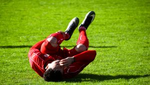
So how does an ACL tear happen? An ACL tear can occur when the knee twists or if the lower leg over extends such as when changing direction rapidly or unexpectedly (like a football or rugby tackle), stopping suddenly or a sometimes even a bad landing. This can result in extreme pain, an unstable knee and reduced range of motion.
How do we go about treating this? Not every patient with an ACL tear will need surgery. Depending of the severity of the injury and the lifestyle of the patient a tear can be managed conservatively with rest and physical therapy. That said, those individuals with an active lifestyle, who do lots of sport or have a severe ACL tear may need reconstructive surgery. Unfortunately, the ligament can’t simply be stitched back together. So, what we have to do it use a graft. We take a bit of ligament from the hamstring or patella tendon and once we have removed all the old torn ACL we replace it with the grafted tendon. In most cases this is done through tiny keyhole incisions.
Whilst success rates for this are high the surgery, like all surgeries, comes with risks including blot clots, infection, pain and stiffness. A full recovery can be 6 months with some individuals taking up to a year as recovery is a very individual thing and it will usually take about 9 months to return to contact sports.

We hope you found this post insightful and interesting…keep an eye on the blog for plenty more upcoming content!
This article is intended to inform and give insight but not treat, diagnose or replace the advice of a doctor. Always seek medical advice with any questions regarding a medical condition.
![]()
Osteoarthritis is a very common type of arthritis – there are many types of arthritis though. It is a condition which gets worse over time or with age which is called degenerative. Although we do see arthritis in younger people it’s often linked to an injury rather than joint wear and tear over time.
The hip joint is a ball and socket joint made up of:
- Bones that form the joints (the femur and the acetabulum)
- Cartilage – a smooth, firm tissue at the ends of the bones
- Joint fluid to lubricate the joints
- Tendons and ligaments to stabilise the joint
- Muscles for movement
In osteoarthritis it is the cartilage at the joint which wears down and essentially doesn’t do its job very well. If this happens the hip joint becomes painful, swollen and stiff. Often this linked to aging but can also be attributed to trauma, fracture, dislocation and in some cases may be hereditary.
Hip arthritis can have varying symptoms which can include:
- Problems with walking
- Pain and stiffness
- Reduced range of movement (usually linked to pain)
- Clicking or creaking sound
- Swelling
How could we treat hip arthritis?
Although there is no true cure for arthritis there are things that can be done to help ease the pain and improve the movement of the joint. These range from conservative manag
ement which includes exercises, resting the joint, medication, physical therapy and, if applicable, losing excess weight. If these fail to have any effect there are surgical options which your consultant would discuss and talk through the range of options available.

We hope you found this post insightful and interesting…keep an eye on the blog for plenty more upcoming content!
This article is intended to inform and give insight but not treat, diagnose or replace the advice of a doctor. Always seek medical advice with any questions regarding a medical condition.
![]()
A kneecap or patella dislocation can be incredibly painful. The kneecap usually sits straight over the knee joint at the front. A dislocation of the patella happens when the knee cap comes out of the groove it sits in on the end of the femur (thigh bone) and ends up on the outside of the knee joint.
A first-time patella dislocation usually happens as a result of a significant injury but the kneecap is more prone to displacement following the initial dislocation. This is because when the patella is dislocated in the first instance the soft tissues (ligaments and tendons) holding it in place are stretched or torn meaning they are less supportive. The most important one of these ligaments is the MPFL (medial patellofemoral ligament). When this ligament tears (which is does in a patella dislocation) it often can heal but not with the correct tension which means that the kneecap is unstable and can pop out of position more easily – recurrent dislocation.
In some people there can be other underlying causes for the knee cap to dislocate repeatedly, like malalignment of the knee joint or the groove not being well developed, which would need to be corrected surgically for successful treatment.
A dislocated kneecap can be fairly obvious as the dislocation causes significant pain and deformity of the knee joint – it will look very out of place and at an unusual angle. Other symptoms can include:
- A painful popping sensation
- Unable to walk
- Unable to straighten the knee
- Sudden swelling of the knee joint especially the front
It is important not to confuse a patella dislocation with a knee joint dislocation which is where the femur or thigh bone displaces from the tibia (shin bone).
How do you treat a patella dislocation?
Often a kneecap dislocation will pop back in by itself, but it is still worth getting it checked by a doctor if you think you have suffered a patella dislocation. Sometimes it won’t pop back into place itself do not try to put it back yourself as this can cause more harm – at this point it is best to phone an ambulance and get it looked at by the hospital doctors.
Once the kneecap has been put back in place by the doctors the treatment can start. This begins as conservative treatment with rest, a brace, crutches and then, once the swelling has gone down, physical therapy.
As previously mentioned a high percentage of first time patella dislocations will progress to recurrent dislocations. At this point there are surgical options available. These can include:
- MPFL reconstruction
- Tibial tubercle transfer
- Corrective osteotomy
We hope you found this post insightful and interesting…keep an eye on the blog for plenty more upcoming content!
This article is intended to inform and give insight but not treat, diagnose or replace the advice of a doctor. Always seek medical advice with any questions regarding a medical condition.
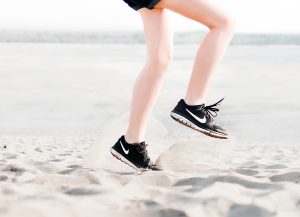
Tendonitis is an injury of the tendon often associated with sudden, explosive movements, such as jumping or throwing, or overuse for example in runners. Outside of sport it can be linked to performing the same movement repeatedly which could be as simple as using a mouse or typing.

Symptoms:
Tendonitis can affect several areas of the body so it is hard to pin down specific symptoms for each area however the following are often common to most areas:
- Pain (particularly increasing on movement)
- Weakness in the area
- Lack of mobility at the joint
- Swelling and sometimes heat
- A grating or grinding feeling as the tendon moves
Often tendonitis will clear up of its own accord with some rest. Initially, stop the activity which caused the tendonitis in the first place. It is important to rest the tendon and the area around it. Then once the symptoms have died down gradually returning to normal activities should be fine. Taking medication such as Ibuprofen to help with the pain and swelling as well as icing the affected area periodically throughout the day may also aid recovery.

Although tendonitis shouldn’t need medical attention if symptoms persist beyond a couple of weeks it is always a good idea to see a GP.
This article is intended to inform and give insight but not treat, diagnose or replace the advice of a doctor. Always seek medical advice with any questions regarding a medical condition.
Upper Limb
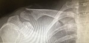
![]()
A broken collarbone or Clavicle is one of the most common fractures in adults. Making up about 5% of all adult fractures a clavicle fracture usually occurs as a result of falling on the shoulder of an outstretched arm. These are often associated with cycling injuries, car collisions or slips and falls.
The clavicle is found between the ribcage (sternum) and the shoulder blade (scapula) and acts to join the arm to the rest of the body.

As mentioned, clavicle fractures are quite common but can vary in type and location. Most fractures occur in the middle part of the bone (the shaft of the bone). Other cases may see the bone break where it attaches to the rib cage or the shoulder blade. The fracture may be a single break or many pieces and could be lined up or far out of place.
A clavicle fracture can be very painful making it hard to move the arms. It is also characterised by downward sagging of the shoulder, a bump or deformity over the break and bruising, swelling and tenderness over the collarbone.
The doctor will ask how the injury occurred and how the shoulder feels. There is usually a bump of bone at the fracture site but an x-ray will confirm the precise location and extent of the fracture.
Treatment:
- Supporting the arm: A simple sling can help support the arm and aid comfort immediately after the break. This can hold the arm in place whilst the bone heals.
- Pain medication: Medication can be used to help manage the pain of a collarbone fracture.
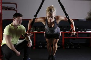
- Physical therapy: Once the injury is healed, elbow and shoulder mobility exercises can be done to prevent stiffness.
- Surgical treatment: If the broken parts of the bone are significantly out of line surgery may be recommended.
- Open reduction and fixation: Plates and screws can be used to hold the broken fragments in the correct place until healing has taken place. Usually these plates and screws will stay in for life unless they are causing discomfort.
- Sometimes a long pin can be used instead of plates and screws to hold the bones in place whilst they heal. This pin usually goes long ways inside the bone from one end across the site of the break.
After surgery there may be some pain, this is part of the natural healing process and simple pain relief medication can help relieve this. Once the fracture is healed physical therapy can help restore full range of movement and strength in the shoulder.
It can take several months for a full recovery from a clavicle fracture. Most people return to regular activities within three months of the injury but the doctor will advise when the injury is stable enough to return to normal activities.
This article is intended to inform and give insight but not treat, diagnose or replace the advice of a doctor. Always seek medical advice with any questions regarding a medical condition.
![]()
A frozen shoulder, also called adhesive capsulitis, is a condition which presents with stiffness and pain in the shoulder joint.
What are the symptoms of a frozen shoulder?
Generally, the condition will develop slowly, in a staged process, and resolve itself over time.
- Initially the freezing stage sees pain on moving the shoulder with the shoulder joint then becoming stiff, limiting the range of movement.
- Then the frozen stage follows whereby the pain begins to subside however the shoulder joint is now stiffer making movement and use more troublesome.
- Finally, the thawing stage, sees range of movement begin to return to the joint and the shoulder begins to improve.

What can cause a frozen shoulder?
The shoulder joint is made up of bones, ligaments and tendons which are all encased by a capsule ligament made up of connective tissue. It is this capsule ligament which can be blamed for a frozen shoulder. The ligament becomes thicker and tightens around the shoulder joint leading to a reduction in movement. Sometimes the cause of this thickening is unknown whilst the chance of the ligament becoming problematic is increased in individuals with diabetes or if they have had to immobilise the shoulder for a prolonged period (e.g. after surgery).
Are there risk factors?
There are certain factors which may increase the risk of developing a frozen shoulder. These can include:
- Age – individuals over the age of 40 tend to be more susceptible to a frozen shoulder
- Immobility – individuals with reduced mobility at the shoulder are at a higher risk. This could include recovery from surgery, broken arm, stroke, rotator cuff tear.
- Systemic diseases – some diseases may predispose individuals to being at risk of a frozen shoulder. These can include diabetes, over or under active thyroid, cardiovascular disease, tuberculosis and Parkinson’s disease.

How do you treat a frozen shoulder?
Usually, frozen shoulder will right itself. It may take a few months, even up to 18, but mostly they do get better on their own. It the problem persists or becomes unmanageable there are options.
- Medications – pain relief and anti-inflammatories such as ibuprofen can help with the pain of a frozen shoulder.
- Physical therapies – range of motion exercises can help improve and restore shoulder mobility.
- Steroid injections – an injection of corticosteroids into the shoulder can help control the pain to aid increased mobility whilst the shoulder gets back to normal.
- Joint distension – an injection of sterile water into the joint capsule can lubricate the joint and help ease up the movement.
- Shoulder manipulation – whilst under general anaesthetic the shoulder is manipulated through the range of movement to loosen the tightened ligament. This is done under anaesthetic so no pain is felt during the manipulation.
- Surgery – whilst surgery for a frozen shoulder is a last resort, if nothing else has helped and symptoms are still progressing, scar tissue and adhesions inside the joint can be removed surgically.
This article is intended to inform and give insight but not treat, diagnose or replace the advice of a doctor. Always seek medical advice with any questions regarding a medical condition.
![]()
A rotator cuff tear is a common injury and cause of pain. It will lead to a weak shoulder making daily activities including brushing hair or putting on jumpers tricky.
The shoulder joint is made up of:
- Bones – humerus (upper arm bone), scapula (shoulder blade) and clavicle (collar bone). The humerus and scapula make up a ball and socket joint where the top of the humerus fits into a shallow socket in the scapula.
- Muscles – the rotator cuff muscles (there are four of them) hold the arm in the shoulder socket. The muscles all come together as tendons to make a covering around the humerus. By doing this it holds the humerus to the shoulder blade allowing the arm to be lifted and rotated.
- Bursa – this is a lubricating sack between the rotator cuff and the bone on the top of the shoulder which allows the rotator cuff tendons to move easily.
If one or more of the rotator cuff tendons is damaged the attachment of the muscle to the bone is compromised.
- Partial tear: this is an incomplete tear and damages the tendon whilst not completely severing it, like a fray.
- Full tear: this is a complete tear where the tendon separates from the bone, there is basically a hole in the tendon.

Why do we get rotator cuff tears?
- Acute: this is usually due to trauma or over straining for example, falling on an outstretched arm or lifting something too heavy with jerking action.
- Degenerative: this is where the tendon becomes worn down over time. This does happen with aging and usually occurs in the dominant arm. There are risk factors associated with this type of tear:
- Bone spurs: Bony spurs can occur as we age, these are overgrowth of bone which when the arms are lifted can rub on the rotator cuff tendon and weaken it.
- Repeated motions: The same movement over and over can place excess stress on the rotator cuff muscles and tendons, this is often seen in sports players (tennis, rowing etc.).
- Lack of blood supply: The blood supply to the rotator cuff can become diminished. In order to repair any damage, the tendon needs a blood supply. This therefore leaves the tendon more at risk of tear.
What does a rotator cuff tear feel like?
The most common symptoms can include:
- Pain – at rest, at night, if lying on the area, when lifting, lowering or rotating the arm.
- Weakness when lifting or rotating the arm.
- Crepitus (crackling sensation in the shoulder).
Acute tears may be associated with a snapping sensation accompanied with immediate arm weakness as these are often due to trauma.
Degenerative tears tend to be more associated with a build up of pain and weakness overtime as the tendon gradually breaks down.

How do we treat rotator cuff tears?
The vast majority of patients can be treated non-surgically to relieve pain and improve shoulder function. This can include:
- Rest: Reducing the movement at the shoulder whilst the injury heals.
- Altering activity: Avoiding activities which aggravate shoulder pain.
- Medication: Anti-inflammatory drugs for example ibuprofen to help reduce swelling and pain.
- Physical therapy: Exercises to promote movement and strengthen the shoulder to increase stability of the shoulder, reduce pain and prevent further injury.
- Injection: If other non-invasive treatments do no relieve pain, an injection may help. It contains local anaesthetic and an anti-inflammatory medicine.
If pain does not improve with non-surgical treatments, your consultant may suggest a surgical treatment. Surgery aims to repair the tear by re-attaching the tendon to the head of the humerus. Although if it is a partial tear may only need trimming or smoothing (called debridement). Many surgical repairs can be done as a day case so there is no need to spend the night in hospital.
There are essentially three methods for repairing a rotator cuff tear:
- Open repair: Traditional open surgery – this is sometimes used if the tear is large and complex.
- Arthroscopic repair: The surgeon makes a small incision and completes the procedure guided by a camera. This is considered minimally invasive surgery.
- Mini-open repair: This is a combination of the other two techniques.
The majority of patients report a reduction in pain and an improvement in shoulder strength following repair surgery. A full recovery can take several months and is enhanced with a tailored exercise programme and physical therapy.
We hope you found this post insightful and interesting…keep an eye on the blog for plenty more upcoming content!
This article is intended to inform and give insight but not treat, diagnose or replace the advice of a doctor. Always seek medical advice with any questions regarding a medical condition.
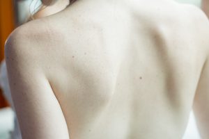
![]()
Shoulder arthritis, specifically osteoarthritis, is a condition which gets worse over time or with age which is called degenerative. Although we do see arthritis in younger people it’s often linked to an injury rather than joint wear and tear.
The shoulder joint is made up of:
- Bones that form the joints (acromioclavicular joint and glenohumeral joint)
- Cartilage – a smooth, firm tissue at the ends of the bones
- Joint fluid to lubricate the joints
- Tendons and ligaments to stabilise the joint
- Muscles for movement
In osteoarthritis it is the cartilage at the joint (usually the acromioclavicular joint) which wears down and becomes less effective. This means that the shoulder can become painful, swollen and stiff. Often this linked to aging but can also be attributed to trauma, fracture, dislocation and in some cases may be hereditary.
Arthritis in the shoulder often presents with pain, particularly when moving or just after moving it. Other symptoms can include reduced range of movement (usually linked to pain) and sometimes even a clicking or creaking sound.
So, how do we go about treating arthritis of the shoulder? First of all, we will try conservative treatments. These can include: rest, heat/ice treatment, physical therapy, range of motion exercises and sometimes medication can help (NSAID – Non-Steroidal Anti-Inflammatory Drugs). If conservative management fails to help we can look at surgical options which include: decompression surgery, shoulder joint replacement or humeral head replacement. Each patient is different so the consultant will help to choose the right option for the individual patient. x

We hope you found this post insightful and interesting…keep an eye on the blog for plenty more upcoming content!
This article is intended to inform and give insight but not treat, diagnose or replace the advice of a doctor. Always seek medical advice with any questions regarding a medical condition.
![]()
What is carpal tunnel syndrome?
Carpal tunnel syndrome (CTS) is the commonest form of nerve entrapment and occurs when the median nerve is compressed at the wrist in the carpal tunnel. It needs timely treatment to prevent avoidable, irreversible and disabling loss of feeling and power in the hand.
The median nerve and finger bending tendons pass through a tunnel across the front of the wrist. The tunnel is made of wrist bones covering the floor and walls and the roof is made of a strong fibrous ligament. Normally the pressure in the canal is very low. The increase in pressure in the tunnel could compress the nerve, compromise the blood supply to the nerve and cause pain/numbness and tingling.
What are the symptoms of CTS?
This is usually felt as pain, tingling or numbness in part of the hand, usually the thumb, index and middle fingers. Sometimes the symptoms are felt in the whole hand. The intensity of pain, tingling and numbness are often quite variable in severity. The symptoms are typically worse at night and with certain daytime activities such as driving, cycling and reading. Shaking the hand may relieve the symptoms. One or both hands can be affected.
Why does it occur?
In most cases, it’s not known what causes the median nerve to become compressed, although the following increase the risk of developing CTS.
- Family history
- Health conditions – diabetes, an underactive thyroid gland or rheumatoid arthritis and obesity
- Pregnancy – Many cases resolve after the baby is born.
- Injuries to the wrist
- Certain activities
- Playing a musical instrument
- Assembly packing
- Work that involves manual labour
- Work with vibrating tools, such as chainsaws
- Other
- Cysts, growths or swellings in the carpal tunnel
- Some drugs used to treat breast cancer – such as exemestane (Aromasin)

What happens if nothing is done?
As carpal tunnel syndrome progresses, you may feel you are losing coordinated movement of your fingers. You may notice weakness in your fingers that makes it hard to do daily tasks, such as buttoning buttons. The symptoms of pain/numbness will increase and most people are particularly troubled by night waking often in the early hours of the morning. If left for too long permanent numbness and muscle weakness could reduce the hand function considerably.
Investigations:
How is it diagnosed?
Carpal tunnel syndrome is diagnosed by a hand specialist based on an evaluation of your symptoms, physical examination and if needed an electrophysiologic test. The surgeon will also examine the patient‘s neck as this can give numbness and tingling in the hands similar to CTS.
Nerve conduction study/Neurophysiology/EMG – This is performed at a later date by a specialist (Neurophysiologist). Mild electric shocks are sent up and down the arm and the strength and speed of their conduction is measured. The test measures the electrical signals passing through the median nerve and how the muscles that are supplied by the nerve are being affected. It can show if the nerve is compressed. Like all tests it is not completely reliable so it can be normal although the patient has CTS and it can be abnormal when the patient does not have CTS. Your Hand specialist will interpret the results in the light of the previous description of symptoms and examination and advise you accordingly.
Rarely X-rays, Ultrasound/MRI scans, blood tests are performed to rule out other causes of CTS
Treatment:
What are the non-operative treatments?
Wrist splint is often the first step in treatment. A splint can be particularly helpful for people whose symptoms are bothering them at night. Wearing a splint during the night can keep the wrist from bending or being extended while you sleep. That often relieves the nerve pressure and eliminates nighttime discomfort. Wearing a splint during the day also can be useful if symptoms persist during waking hours. Some people don’t like wearing a splint during the day, though, because moving the wrist naturally may be difficult.
Corticosteroid injection into the carpal tunnel may help reduce symptoms.
- Corticosteroids lower inflammation and swelling within the carpal tunnel, which can relieve pressure on the median nerve.
- Benefit may be temporary.
- Rarely more than two injections are beneficial
- If the symptoms are marked and established, surgery may be appropriate
- Where the clinical picture is unclear a positive response to an injection helps confirm the diagnosis.
What does the operation involve?
The operation is called a carpal tunnel release (CTR) or decompression (CTD). The operation is usually performed as a day case surgery (means, you won’t have to stay in hospital overnight.) under local anaesthetic.
On the day of surgery
A local anaesthetic is used to numb your hand and wrist, but you’ll remain awake throughout the operation. General anaesthetic is rarely advised (patients with needle phobia). A tourniquet, like a blood pressure cuff, is placed around the top of the arm. It is inflated (tightened) during the operation to reduce bleeding, which makes the operation easier and safer. It can be a little uncomfortable, but is almost always well tolerated by our patients. Before the surgery, the arm is painted with an antiseptic and covered with sterile sheets, to help minimise the risk of infection.
The surgeon checks the skin for numbness and if needed injects more local anaesthetic. Once the skin becomes numb, the surgeon makes a cut over the front of the wrist, and the roof over the tunnel is released, so that the nerve has more space in its tunnel. The skin is then stitched up usually with stitches. A supportive dressing is applied and the patient’s arm(s) elevated.
The total time in hospital is usually 1-2 hours. For most hand conditions surgeons avoid operating on both hands at once as it can be significantly disabling. The decision to release both sides at once is a decision between the patient and the surgeon.

What happens after surgery?
The care of the hand in the post-operative period is very important in helping to achieve a good result. The local anaesthetic wears off 4-10 hrs after surgery, patients are encouraged to start taking painkillers before the pain starts i.e. on return home and for at least 24 hours from there. This way most of our patients report little or any pain. Initially the aims are comfort and elevation of the hand in a bandage for a couple of days, and you may need to wear a sling. Gently exercise your fingers, shoulder and elbow to help prevent stiffness. You may be able to start these gentle exercises on the day of your operation.
After having surgery for CTS, you can use your hand to do light activities that don’t cause excessive pain or discomfort. Try to avoid using your hand for more demanding activities until it’s completely recovered, which may take several weeks.
The bandage can be removed after 2-7 days, leaving a sticky dressing beneath. The patient or GP practice nurse can do this. The patient is reviewed in clinic around 2 weeks following the operation for wound check and removal of sutures.
The wound should be massaged (typically after 2 weeks) using moisturising cream by the patient 3 times a day for 3 months once the wound is well healed. This reduces the scar sensitivity and scar related complications (tenderness, helps scar to mature). Hand therapy for scar management will be needed in few patients.
Most patients can drive after the wound has healed (usually 2 weeks). Most patients return to work in 2-3 weeks, but this varies with occupation; heavy manual work usually takes about 6 weeks. Patients should avoid pressing heavily on the scar for 3 months following the operation as this will be quite painful.

Are there any risks?
Any operation can have unforeseen consequences and leave a patient worse than before surgery. For CTD the risks are small but include:
- The scar may be tender in about 20% of patients. This usually improves with scar massage, over 3 months.
- Aching, especially on gripping. This occurs in about 4% of patients and also improves with time. Grip strength can also take some months to return to normal.
- Stiffness may occur in particular in the fingers. This is usually short-term and only infrequently requires physiotherapy. But it is very important that it is resolved quickly to avoid permanent stiffness. This occurs rarely but can do associated with CRPS (see below).
- Numbness over the base of the thumb, caused by damage to a branch of the nerve, happens in less than 4% of patients. This rarely causes any functional problems.
- Wound infections occur in about 1% of cases. These usually quickly resolve with antibiotics.
- Chronic Regional Pain Syndrome “CRPS”. This is a rare but serious complication, with no known cause or proven treatment. The nerves in the hand “over-react”, causing swelling, pain, discolouration and stiffness, which very slowly improve.
- Injury to the main median nerve can occur extremely rarely, resulting in permanent numbness and weakness in the hand.
- Failure to fully separate the roof of the carpal tunnel during surgery, usually resulting in persistent CTS symptoms
- In rare cases, the return of CTS symptoms long after apparently successful surgery
What are the results of the operation?
The overall success rate of carpal tunnel open surgery is more than 95% with a complication rate of less than 3%. Most patients have very rapid or immediate relief of their pain. Symptoms of numbness or weakness may well never resolve particularly if there was continuous numbness or weakness prior to surgery (permanent damage to the nerve even before the surgery). Nonetheless most patients gain significant benefit in these symptoms, which may improve for up to 2 years from surgery.
Is surgery a permanent cure?
Yes, in majority of patients. However the reported recurrence rates after carpal tunnel decompression are between 0.3 and 12%.
![]()
Tennis elbow: What is it and why do we get it?
Tennis elbow is a condition giving rise to pain around the outside of the elbow joint. Its’ official title is lateral epicondylitis. It is a very common condition affecting almost up to 1 in 3 individuals. Golfers elbow however affects the inside edge of the elbow rather than the outside and is known as medial epicondylitis.
The elbow joint is made up of:
- Bones – ulna, radius (lower arm) and humerus (upper arm)
- Ligaments (join bones to bones)
- Tendons (attach muscles to bones)
- Muscles
Some of the muscles that create bending and straightening of the forearm and wrist start from the same tendon. When these muscles are used repeatedly under force (such as a sportsperson) it can cause an overuse strain of this common tendon. That said, tennis elbow can also affect other muscles and tendons around the elbow. When this strain occurs in the tendons and muscles it can lead to inflammation and small tears. Although the name suggests that this injury is common to tennis players and sports players it is also seen in other professions involving repeated strain of the elbow including violin players, decorators and window cleaners!

Treatment options:
Usually tennis elbow will although it might take longer than one would like. There are treatment options available which may alleviate symptoms and speed up the recovery process. To help tennis elbow get better it is crucial to rest the elbow so stopping the activity which has led to the problem for a while until it is healed. Although that might be easier said than done it may slow down the recovery significantly if the joint isn’t rested. Icing the joint can also help to alleviate symptoms of tennis elbow.

We hope you found this post insightful and interesting…keep an eye on the blog for plenty more upcoming content!
This article is intended to inform and give insight but not treat, diagnose or replace the advice of a doctor. Always seek medical advice with any questions regarding a medical condition.
Within the UK, NICE (National Institute for Health and Clinical Excellence) issues clinical guidelines for healthcare treatments. These treatments must be backed by clinical evidence and plenty of research to be approved by NICE. Shockwave Therapy is supported by NICE guidelines for certain conditions with more indications for use being added each year.
But what actually is Shockwave Therapy? It is a treatment which can be used in long term soft tissue injuries, initially it was used in the treatment of Tendinopathies, and some specific bone conditions. The nice thing about Shockwave Therapy is that the side effects are minimal. The therapy uses high energy sound wave pulses which is totally non-invasive – there is no skin incision. These sound waves are produced by a hand-held device which is incredibly accurate. The sound waves cause increased blood flow to the problem area which helps promote healing of that area for example a painful tendon. It is important to note that it is not the sound waves themselves but the resulting increased blood flow which has the positive effect on the damaged or painful tissue. It is thought that this improved blood flow delivers more nutrients to the damaged area speeding up the natural rate of healing.
What is Shockwave Therapy used for?
Shockwave Therapy can be effective when used to treat a range of soft tissue and bony conditions particularly when scar tissue is contributing to the pain. These can include (not limited to):
- Shoulder pain
- Tennis elbow
- Golfer’s elbow
- Tendonitis
- Heel pain
- Plantar fasciitis
Why does it work well when there is scar tissue?
Scar tissue by nature is non-elastic and quite tough whereas the body’s normal soft tissue is elastic and springy. For this reason, if a tendon (attaches muscle to bone so needs to be springy) becomes scarred it may not work as effectively as when it was a healthy tendon. This reduced function can then affect surrounding tissues as well leading to further pain. By breaking down the scar tissue (through shockwave therapy) the affected area can then regenerate healthy tissue and return to normal function.
It is important to note that no one treatment works for every patient so it is vital to discuss and explore all the options available.
We hope you found this post insightful and interesting…keep an eye on the blog for plenty more upcoming content!
This article is intended to inform and give insight but not treat, diagnose or replace the advice of a doctor. Always seek medical advice with any questions regarding a medical condition.
![]()
Subacromial impingement
Subacromial impingement is one of the most common shoulder injuries and it develops as a result of the should tendon being caught or rubbing on the bone as the arm is lifted. More often than not this condition affects the rotator cuff tendon. This is the elastic tissue which connects the shoulder muscles to the upper arm bone. Whilst it is the tendon catching on the bone which causes the pain, the reason this happens in the first place can be more complex.
So what happens?
As the arm is lifted, the rotator cuff tendon passes through a narrow passage (called subacromial space). It is the bone at the top of this passageway which the tendon catches on to cause the pain. Under normal circumstances the bone would not catch the tendon but sometimes one of the following can happen causing the tendon to catch on the bone:
- The area around where the tendon joins the bone (called bursa) can become inflamed and irritated
- The whole tendon become inflamed, thickened or swollen due to overuse injury
- Bone spurs develop to make the passageway narrower
- The bone is curved rather than flat (usually individuals are born with this rather than it developing)
Usually the shoulder impingement will rectify itself with some simple exercised but on occasion can drag on and become more of a problem.
What does subacromial or shoulder impingement feel like?
- Pain – in the top and outside of the shoulder
- Pain – worsening when the arm is lifted
- Pain and aching – worsening at night
- Arm weakness
- But it won’t be still
When to seek medical help?
If the pain doesn’t rectify itself after a couple of weeks and is preventing normal daily activities being performed it could be advisable to see one of us or a GP.
What can you do to help?
- Stop doing the activities which make the pain worse (usually lifting the arm above the head repeatedly) for a couple of days or weeks until it heals.
- Keep the arm mobile – it should prevent the shoulder becoming stiff or weak.
- Icing the shoulder for 20 minutes a few times per day can help (not directly on the skin though).
- Simple painkillers (ibuprofen or paracetamol) may also help.
- A physiotherapist may give some exercise to keep the shoulder mobile whilst it heals.
- Usually these will rectify a shoulder impingement but in some cases further treatment might be needed.
We hope you found this post insightful and interesting…keep an eye on the blog for plenty more upcoming content!
This article is intended to inform and give insight but not treat, diagnose or replace the advice of a doctor. Always seek medical advice with any questions regarding a medical condition.